Biology - 2024
Complete Detailed Solutions · Previous Year Paper
Group A — Very Short Answer Type
1. (I) The portion of the growth curve where rapid growth of bacteria is observed is known as ____________
Log phase or Exponential phase.
1. (II) The human eye can focus objects at different distances by adjusting the focal length of the eye lens. This is due to__________.
Accommodation (specifically, the action of the ciliary muscles changing the lens curvature).
1. (III) What is meant by the term osmoregulation?
Osmoregulation is the active regulation of the osmotic pressure of an organism's body fluids to maintain the homeostasis of the organism's water content (maintaining fluid and electrolyte balance).
1. (IV) Who is known as father of genetics?
Gregor Johann Mendel.
1. (V) What is meant by power of accommodation of the eye?
The power of accommodation is the ability of the eye lens to automatically adjust its focal length to form a sharp image on the retina of objects situated at varying distances.
1. (VI) Enzyme increases the rate of reaction by lowering the activation energy. Is this statement true or false?
True.
1. (VII) What is cistron?
A cistron is a section of a DNA or RNA molecule that codes for a specific polypeptide in protein synthesis; it is essentially the functional unit of a gene.
1. (VIII) State two economically important uses of heterotrophic bacteria.
1. Dairy Industry: Lactobacillus is used to convert milk into curd/cheese.
2. Pharmaceuticals: Used in the production of antibiotics (e.g., Streptomyces producing streptomycin).
1. (IX) What is the main function of kinase?
The main function of a kinase enzyme is to catalyze the transfer of a phosphate group from a high-energy molecule (like ATP) to a specific substrate (a process called phosphorylation).
1. (X) The growth of bacterial population follows a geometric progression. True or False?
True (Bacteria divide by binary fission, doubling their population each generation, which is a geometric/exponential progression).
1. (XI) Are viruses living or non-living?
Viruses are considered to be at the boundary between living and non-living. They are non-living (inert particles) outside a host cell, but exhibit living characteristics (replication and genetic mutation) when they infect a living host cell.
1. (XII) Fluid Thioglycollate medium is used for the cultivation of which type of organism?
It is used primarily for the cultivation of anaerobic bacteria (as well as microaerophiles and aerobes, to determine their exact oxygen requirements based on where they grow in the tube).
Group B — Short Answer Type
2. How are co-factors different from prosthetic groups?
Both are non-protein components essential for the catalytic activity of certain enzymes (holoenzymes), but they differ in how they bind to the enzyme:
- Co-factors: This is a broad term for non-protein helper molecules. Specifically, when referring to inorganic ions (like Mg²⁺, Zn²⁺) or loosely bound organic molecules (coenzymes like NAD⁺), they bind transiently and loosely to the apoenzyme. They usually associate with the enzyme only during the chemical reaction.
- Prosthetic Groups: These are organic or inorganic non-protein molecules that are bound tightly and permanently (often covalently) to the enzyme. An example is the heme group tightly bound to hemoglobin or cytochromes.
3. Give characteristics of genetic code.
The genetic code is the set of rules by which information encoded in genetic material (DNA or mRNA sequences) is translated into proteins. Its key characteristics are:
- Triplet Code: Three consecutive nucleotide bases (a codon) code for one specific amino acid.
- Universal: With few exceptions, the same codons code for the same amino acids in almost all organisms, from bacteria to humans.
- Unambiguous (Specific): A specific codon always codes for only one specific amino acid (e.g., UUU always codes for Phenylalanine).
- Degenerate (Redundant): Multiple different codons can code for the same amino acid (e.g., UUU and UUC both code for Phenylalanine).
- Non-overlapping and Commaless: The code is read continuously, three bases at a time, without any overlapping bases or gaps/commas between codons.
4. Differentiate between DNA/RNA.
| Feature | DNA (Deoxyribonucleic Acid) | RNA (Ribonucleic Acid) |
|---|---|---|
| Sugar | Deoxyribose sugar (lacks one oxygen atom). | Ribose sugar. |
| Nitrogenous Bases | Adenine (A), Guanine (G), Cytosine (C), Thymine (T). | Adenine (A), Guanine (G), Cytosine (C), Uracil (U) instead of Thymine. |
| Structure | Usually Double-stranded (Double Helix). | Usually Single-stranded. |
| Function | Stores and transfers long-term genetic information. | Acts as a messenger (mRNA) to transfer code to ribosomes to make proteins. |
| Stability | Highly stable, less prone to mutation. | Less stable, highly reactive, prone to rapid degradation. |
5. Explain Krebs cycle. draw suitable flowchart for explanation.
The Krebs Cycle (also known as the Citric Acid Cycle or TCA cycle) is a series of chemical reactions used by all aerobic organisms to generate energy through the oxidation of acetyl-CoA derived from carbohydrates, fats, and proteins into carbon dioxide and chemical energy in the form of ATP (or GTP). It occurs in the mitochondrial matrix of eukaryotes.
Flowchart of the Krebs Cycle:
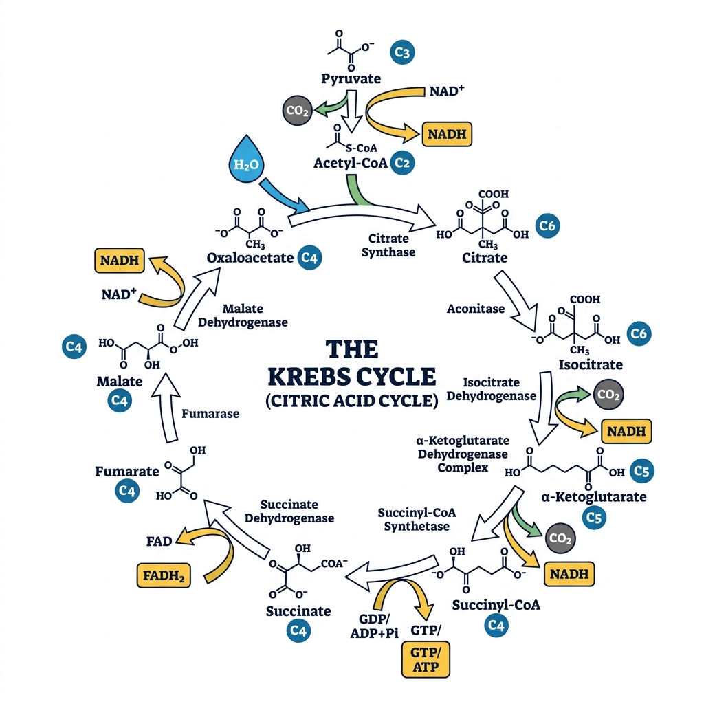
Note: For every molecule of Acetyl-CoA entering the cycle, it produces 2 CO₂, 3 NADH, 1 FADH₂, and 1 ATP/GTP.
6. The sequence of the coding strand of DNA in a transcription unit is mentioned below.
3′ AATGCAGCTATTAGG 5′
Write the sequence for:
1. Its complementary strand
2. Its mRNA
Understanding the strands:
The sequence provided is the Coding Strand (Non-Template strand). The question writes it in the 3' to 5' direction: 3' AATGCAGCTATTAGG 5'. Normally, the coding strand is written 5' to 3', but we must follow the polarity given.
1. Its Complementary Strand (The Template Strand):
The template strand is antiparallel and complementary (A pairs with T, C pairs with G).
Coding: 3' A A T G C A G C T A T T A G G 5'
Template: 5' T T A C G T C G A T A A T C C 3'
2. Its mRNA sequence:
The mRNA sequence is identical to the Coding Strand sequence, except all Thymines (T) are replaced by Uracils (U). The polarity remains exactly the same as the coding strand.
Coding: 3' A A T G C A G C T A T T A G G 5'
mRNA: 3' A A U G C A G C U A U U A G G 5'
(If rewritten in the standard 5' to 3' format, it would be: 5' GGAUUAUCGACGUAA 3')
Group C — Long Answer Type
7. Illustrate the two models by which an enzyme holds the substrate.
Enzymes are highly specific catalysts. The way they bind their specific substrates to their active sites is explained by two primary models:
1. The Lock and Key Model (Emil Fischer, 1894):
This model postulates a rigid binding mechanism.
- Concept: The enzyme's active site is viewed as a rigid "lock," and the substrate is the "key."
- Mechanism: The geometry of the substrate exactly matches the pre-existing, static geometry of the enzyme's active site. They fit together perfectly without any structural changes to the enzyme.
- Limitation: This model explains enzyme specificity well but fails to explain how enzymes stabilize the transition state of a reaction or how non-competitive inhibitors affect the enzyme's shape.
2. The Induced Fit Model (Daniel Koshland, 1958):
This is the more widely accepted, modern model.
- Concept: The enzyme's active site is not a rigid lock, but rather a flexible structure.
- Mechanism: As the substrate approaches and begins to bind, the enzyme undergoes a conformational change (a change in its 3D shape) to mold tightly around the substrate.
- Analogy: It is like a hand entering a glove; the glove is flexible and molds to exactly fit the shape of the hand.
- Advantage: This physical wrapping places strain on the substrate's chemical bonds, lowering the activation energy and perfectly explaining how enzymes stabilize the transition state to catalyze the reaction.
8. Explain steps of Glycolysis in details.
Glycolysis is the first metabolic pathway of cellular respiration, occurring in the cytoplasm of all living cells. It breaks down one molecule of Glucose (a 6-carbon sugar) into two molecules of Pyruvate (a 3-carbon compound), generating a net yield of 2 ATP and 2 NADH. It happens in 10 enzymatic steps, divided into two phases:
Phase I: Energy Investment Phase (Uses 2 ATP)
- Phosphorylation: Glucose is phosphorylated by ATP to form Glucose-6-phosphate (Enzyme: Hexokinase). (-1 ATP)
- Isomerization: Glucose-6-phosphate is rearranged into Fructose-6-phosphate (Enzyme: Phosphoglucose isomerase).
- Phosphorylation: Fructose-6-phosphate is phosphorylated by a second ATP to form Fructose-1,6-bisphosphate (Enzyme: Phosphofructokinase - the main regulatory enzyme). (-1 ATP)
- Cleavage: The 6-carbon Fructose-1,6-bisphosphate is split into two 3-carbon molecules: DHAP and Glyceraldehyde-3-phosphate (G3P).
- Isomerization: DHAP is quickly converted into a second molecule of G3P. From this point on, everything happens twice.
Phase II: Energy Payoff Phase (Generates 4 ATP and 2 NADH)
- Oxidation: The two G3P molecules are oxidized. NAD⁺ is reduced to NADH, and a phosphate group is added, forming two molecules of 1,3-bisphosphoglycerate (1,3-BPG). (+2 NADH)
- ATP Generation: A phosphate group is transferred from 1,3-BPG to ADP, forming two molecules of 3-phosphoglycerate and two ATPs. (+2 ATP)
- Mutase Action: The phosphate group is moved, forming two molecules of 2-phosphoglycerate.
- Dehydration: Water is removed, creating a high-energy double bond in two molecules of Phosphoenolpyruvate (PEP) (Enzyme: Enolase).
- Final ATP Generation: The phosphate group from PEP is transferred to ADP, forming two molecules of Pyruvate and two ATPs (Enzyme: Pyruvate kinase). (+2 ATP)
Net Yield: 4 ATP (produced) - 2 ATP (invested) = 2 ATP. Plus 2 NADH and 2 Pyruvates.
9. Give functions of Proteins as receptors and structural elements.
Proteins are the most versatile macromolecules in living systems. Two of their critical roles are acting as receptors and forming structural elements.
1. Proteins as Receptors (Cell Signaling and Communication):
Receptor proteins are embedded in the cell membrane or found within the cytoplasm. They act as the "eyes and ears" of the cell.
- Signal Reception: They bind to specific extracellular signal molecules (ligands) such as hormones, neurotransmitters, or growth factors. Example: The Insulin Receptor binds insulin in the blood.
- Signal Transduction: Upon binding the ligand, the receptor protein changes its 3D shape. This shape change triggers a cascade of chemical reactions inside the cell, allowing the cell to respond to the outside environment without the signal molecule ever actually entering the cell.
- Examples: G-protein coupled receptors (vision, smell), neurotransmitter receptors (in synapses), and immune system receptors (T-cell receptors that recognize foreign antigens).
2. Proteins as Structural Elements:
Structural proteins provide physical support, shape, and protection to cells, tissues, and entire organisms. They are typically fibrous, tough, and insoluble in water.
- Cytoskeleton: Inside the cell, proteins like actin and tubulin form microfilaments and microtubules. These give the cell its shape, allow it to move, and organize cell division.
- Extracellular Matrix & Connective Tissue: Collagen is the most abundant protein in mammals, forming the structural framework of skin, bones, tendons, and cartilage. Elastin provides elasticity to blood vessels and lungs.
- External Structures: Keratin is the tough structural protein that forms hair, nails, horns, feathers, and the outer layer of human skin, protecting the body from the environment.
10. Describe the characteristics of the individuals with the following chromosomal abnormalities: Trisomy at chromosome 21, XXY, XO.
Chromosomal abnormalities often result from non-disjunction during meiosis, leading to aneuploidy (an abnormal number of chromosomes).
1. Trisomy at Chromosome 21 (Down Syndrome):
- Genotype: 47, XX,+21 or 47, XY,+21 (Three copies of chromosome 21 instead of two).
- Characteristics: Individuals exhibit distinct facial features including a flattened face, upward-slanting eyes, and a short neck. They generally suffer from mild to moderate intellectual disability and developmental delays. They have a higher risk of congenital heart defects, respiratory issues, and early-onset Alzheimer's disease. Muscle hypotonia (low muscle tone) is common in infants.
2. XXY (Klinefelter Syndrome):
- Genotype: 47, XXY (A male born with an extra X chromosome).
- Characteristics: Individuals are phenotypically male but typically have underdeveloped testes and produce lower levels of testosterone. This leads to delayed or incomplete puberty, reduced facial and body hair, and often infertility (azoospermia). They may develop gynecomastia (enlarged breast tissue) and tend to be taller than average with long limbs. Intelligence is usually normal, though some mild learning or language difficulties may be present.
3. XO (Turner Syndrome):
- Genotype: 45, X0 (A female born with only one complete X chromosome, lacking the second sex chromosome).
- Characteristics: Individuals are phenotypically female. They typically have short stature, a webbed neck, a low hairline at the back of the neck, and a broad chest with widely spaced nipples. Crucially, they experience gonadal dysgenesis (streak ovaries), meaning they do not undergo normal puberty, fail to menstruate, and are almost always infertile. Intelligence is generally normal, but there may be deficits in spatial and mathematical reasoning. Congenital heart defects are also common.
11. A tall plant with red flowers (dominant) is crossed with a dwarf plant with white flowers (recessive). Work out a dihybrid cross and state the dihybrid ratio. What will be the effect on the dihybrid ratio if the two genes are interacting with each other?
1. Working out the Dihybrid Cross:
Let the dominant traits be Tall (T) and Red (R). Let the recessive traits be dwarf (t) and white (r).
Parents (P generation): Homozygous Tall Red (TTRR) x Homozygous dwarf white (ttrr)
Gametes: TR x tr
F1 Generation: All offspring are TtRr (Heterozygous Tall and Red).
F1 Selfing (TtRr x TtRr) to get F2 generation:
Each F1 parent can produce four types of gametes: TR, Tr, tR, tr.
We use a 4x4 Punnett Square for the F2 generation:
| TR | Tr | tR | tr | |
|---|---|---|---|---|
| TR | TTRR (Tall, Red) | TTRr (Tall, Red) | TtRR (Tall, Red) | TtRr (Tall, Red) |
| Tr | TTRr (Tall, Red) | TTrr (Tall, White) | TtRr (Tall, Red) | Ttrr (Tall, White) |
| tR | TtRR (Tall, Red) | TtRr (Tall, Red) | ttRR (dwarf, Red) | ttRr (dwarf, Red) |
| tr | TtRr (Tall, Red) | Ttrr (Tall, White) | ttRr (dwarf, Red) | ttrr (dwarf, white) |
Standard Dihybrid Phenotypic Ratio:
- 9 Tall, Red (T_R_)
- 3 Tall, White (T_rr)
- 3 dwarf, Red (ttR_)
- 1 dwarf, white (ttrr)
Ratio = 9 : 3 : 3 : 1
2. Effect of Gene Interaction (Epistasis):
Mendel's 9:3:3:1 ratio assumes the two genes act completely independently of each other. However, if the two genes interact (a phenomenon known as Epistasis, where one gene modifies or masks the expression of the other), the classic 9:3:3:1 ratio will be modified.
For example, if the gene for height was required to express flower color (recessive epistasis), the ratio might become 9:3:4. In dominant epistasis, it could become 12:3:1. In complementary gene interaction, it becomes 9:7. Thus, gene interaction collapses the 4 phenotypic classes into fewer classes, altering the standard Mendelian ratio.